
Respiration: process by which living organisms exchange gases with their environment.
Subprocesses:
- Ventilation: exchange of air into the respiratory organ
- Inhalation
- exhalation
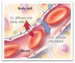
- External respiration : gas exchange between air and blood by diffusion
- internal respiration: gas exchange between blood and tissues, cells
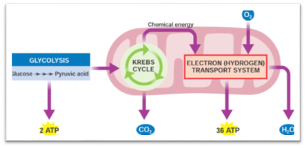
- cellular respiration: biological oxidation
Learn more about the human body's respiratory system.
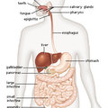
The respiratory tract is the outpocketing of the foregut -> sacklike
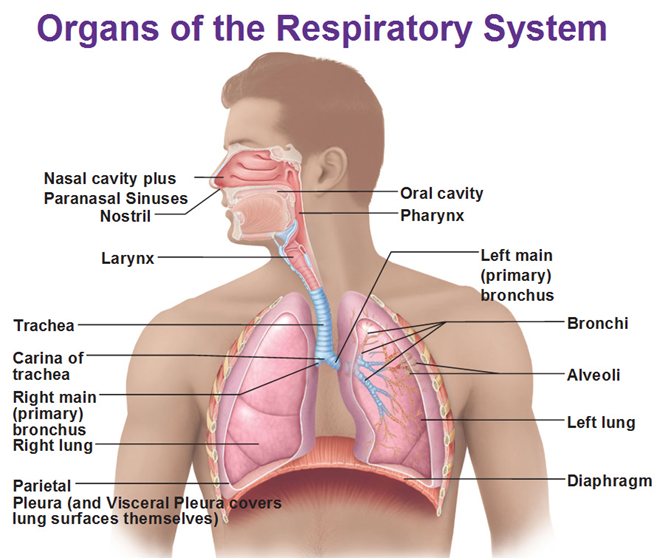
The respiratory system consists of the airways and lungs.
I. Upper airways
- Nostrils -> nasal cavities:
- Mucous membrane with ciliated columnar epithelium
- Glands produce mucus
- Cilia move the mucus towards the pharynx (1-2cm/sec)
- hairs
- rich in capillaries
Function:
- filter air
- warm air
- moisten
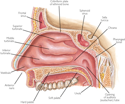
- Pharynx
- where the oral cavity and the nasal cavity meet
- the food and air pathways cross each other
- when you breathe through your mouth, air enters here
- air is not cleaned, warmed up or moistened
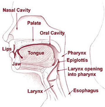
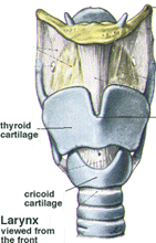
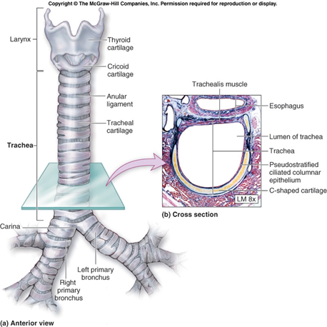
- Larynx = voice box (Adam’s apple)
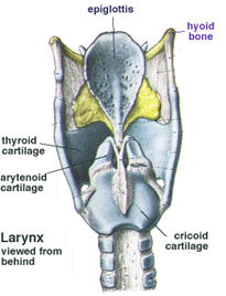
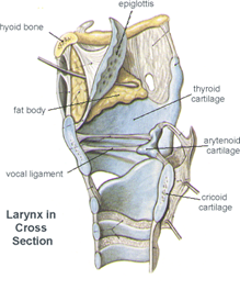
- 1st part of the trachea
- In front of the esophagus
- cartilages:
- thyroid cartilage
- cricoid (=ring-shaped) cartilage
- arytenoid cartilage
- epiglottis
- vocal chords / folds
- infoldings of mucous membrane stretched horizontally across the larynx.
- small skeletal muscles vary the position and tension
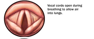
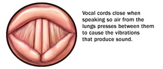
- glottis: the space between the vocal folds
- exhalation -> tense chords vibrate -> sounds + tongue, lips, hard palate => articulated speech
- high pitch sounds: narrow gap, tense chords
- length of vocal fold ~ size of larynx
- children – short
- adolescence – larynx grows -> uncertain voice = mutation
II. Lower airways
4. Trachea
- long tube to the lungs (12cm x 2.5cm)
- ciliated epithelium -> sweeps mucus towards pharynx
- supported by C-shaped cartilage rings -> hold it open
- coughing, sneezing reflex
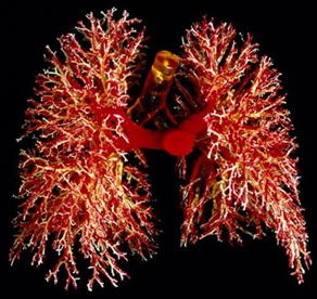
LUNG
- a pair of lungs
- left lung: 2 lobes (heart)
- right lung 3 lobes
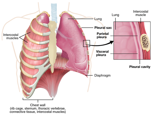
- pleurae cover it
- Double membrane
- Fluid between them
- Stick together, slip past
- Bronchi (bronchus)
- enter the lungs
- have cartilage support, complete rings
- branch repeatedly into smaller airways
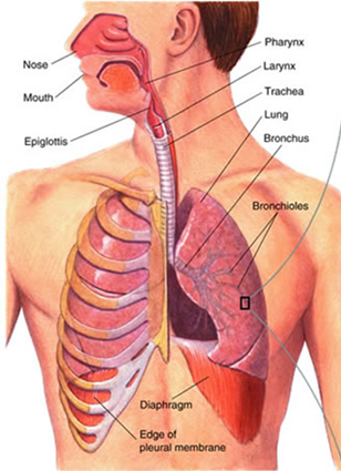
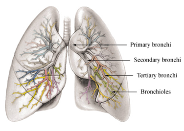
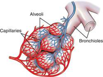
- Bronchioles
- lack cartilage
- smooth muscle
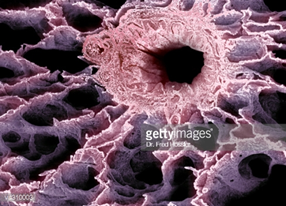
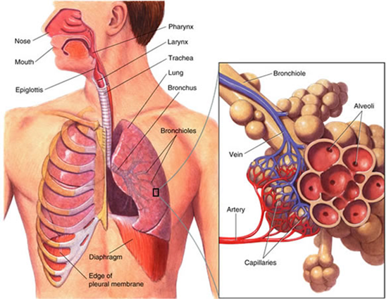
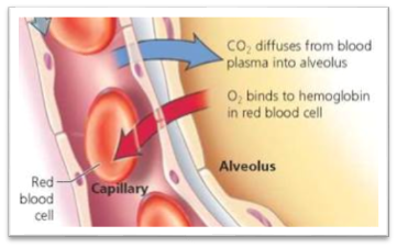
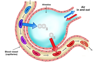
- Alveoli (alveolus)
- air sacks, blind ended cavities
- 600 million
- large respiratory surface ~100m2
- simple squamous epithelium
- surrounded by capillaries, lymph vessels, nerves
- gas exchange:
- gases diffuse across the walls of alveoli and capillaries
- -> blood picks up O2 and gives up CO2
Breathing
Process of filling and emptying internal respiratory organs
Thoracic cavity:
- enclosed by ribs
- separated from the abdominal cavity by the diaphragm
- a sheet of muscles
- domeshaped at rest
- ribs: curve from the backbone, meet at the sternum
- intercostal muscles: diagonal set of muscles between ribs
- pleural membrane:
- double layer
- encloses the lung
- covers the muscles
- thin layer of (pleural) fluid between them -> stick together, slip past
- double layer
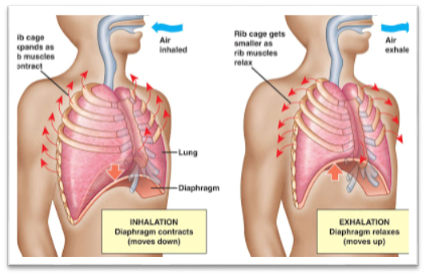
Inhalation:
- active process
- diaphragm contracts -> moves downward towards abdominal cavity
- intercostal muscles contract -> rib cage is raised up and out
- => volume expands -> lowers air pressure slightly below the atmospheric pressure -> suck air in -> lung follows passively!
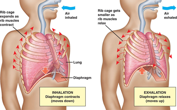
Exhalation:
- passive process at rest
- muscles and diaphragm relax –
- -> volume decreases
- -> pressure rises
- -> expulsion of air
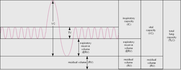
Capacity of the lungs
Breathing volume: 0.5 litre
Inspiratory reserve volume: + 2.5 litres (additional volume of air we can inhale)
Expiratory reserve volume: + 1 litre (additional volume of air we can exhale)
Residual volume: 1.5 l (can’t leave, only if compressed, keeps the lung from collapsing)
Vital capacity : The maxium amount of air that can be exhaled after a forced inhalation.
= breathing volume + inspiratory reserve volume + exspiratory reserve volume (about 4 litres)
Spirometer: an apparatus used for measuring the volume of air inspired and expired by the lungs
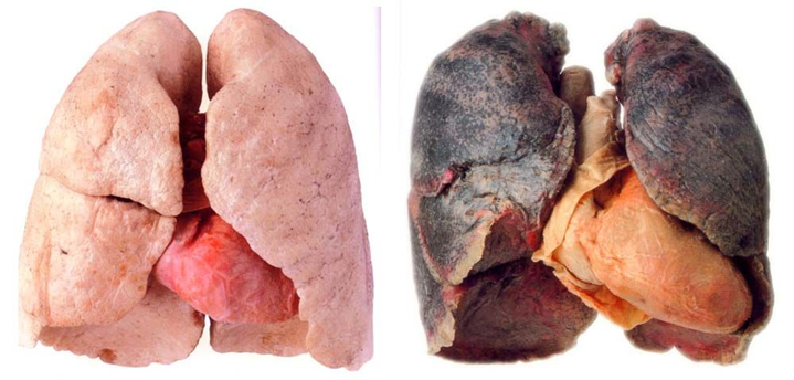
Diseases
1) Bronchitis: an inflammation of the membranes of the bronchial tubes
2) Asthma:
an allergic response characterized by the constriction (narrowing) of the bronchial tubes
can be caused by many different factors
medications and inhalers are used to open up the respiratory passages
3) Emphysema:
a general deterioration of the lungs
alveoli are enlarged and stiff (and can't function) which reduce the lung capacity for air
4) Pneumonia:
the alveoli become filled with fluid, preventing the normal exchange of gases
can be life threatening
5) Lung Cancer:
tumors form in the lungs as a result of irregular and uncontrollable cell growth
The effect of smoking:
Surprising facts about the respiratory system
Ajánlott bejegyzések:

A bejegyzés trackback címe:
Kommentek:
A hozzászólások a vonatkozó jogszabályok értelmében felhasználói tartalomnak minősülnek, értük a szolgáltatás technikai üzemeltetője semmilyen felelősséget nem vállal, azokat nem ellenőrzi. Kifogás esetén forduljon a blog szerkesztőjéhez. Részletek a Felhasználási feltételekben és az adatvédelmi tájékoztatóban.